
Nephroprotective activities of the aqueous seed extract of Carica papaya
Nephroprotective Activities of the Aqueous Seed Extract of Carica papaya Linn. in Carbon Tetrachloride-Induced Renal Injury in Wistar Rats: A Dose- and Time-Dependent Study
Research Article | Biology and Medicine | Volume 1, Issue 1 (2009), Pages 11–19
The kidneys play an essential role in maintaining the body's internal balance by filtering waste products from the blood, regulating fluid and electrolyte levels, and supporting several metabolic processes. However, exposure to environmental chemicals and certain toxic substances can interfere with normal kidney function and lead to tissue damage. Understanding how natural plant extracts may protect renal tissues against toxic injury has therefore become an interesting area of biomedical research.
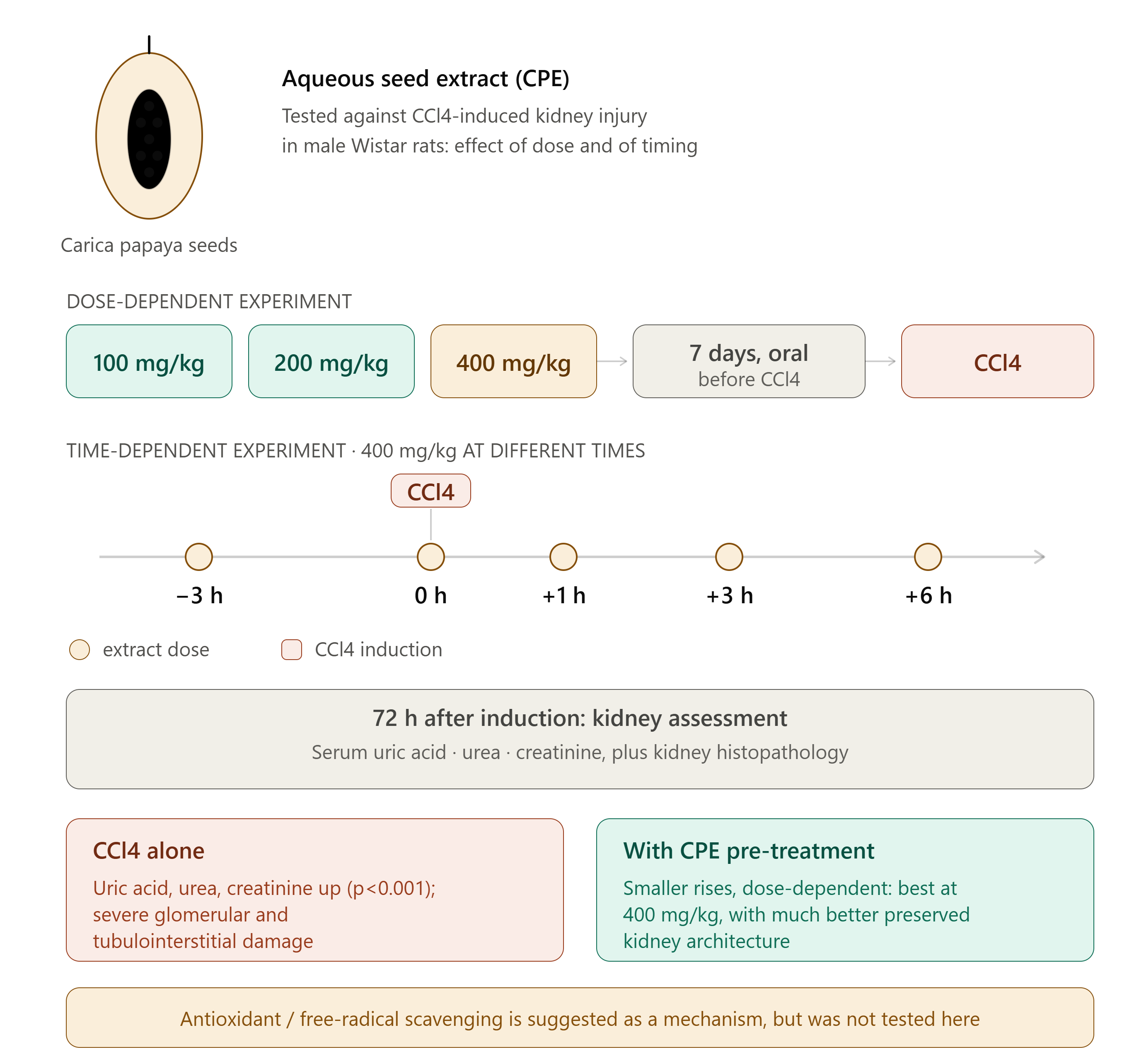
This study investigated the nephroprotective potential of an aqueous seed extract of Carica papaya Linn. (CPE) using a carbon tetrachloride (CCl4)-induced renal injury model in male Wistar rats. Particular attention was given to the influence of extract dosage and administration timing on the protective response.
For the dose-dependent experiment, animals received oral doses of 100, 200 or 400 mg/kg body weight of the aqueous extract for seven consecutive days before exposure to CCl4. A separate experiment evaluated the time-dependent effects of the 400 mg/kg dose, administered at different intervals in relation to CCl4 induction, including three hours before exposure and at 0, 1, 3 and 6 hours after exposure.
Renal function was assessed by measuring serum uric acid, urea and creatinine concentrations 72 hours after the induction of renal injury. In addition, histopathological examinations were performed to evaluate structural changes in kidney tissues.
The administration of CCl4 produced significant increases in serum uric acid, urea and creatinine levels (p<0.001), accompanied by severe histological alterations, particularly glomerular and tubulointerstitial damage. In contrast, pretreatment with the aqueous Carica papaya seed extract reduced the elevations in these biochemical parameters. The protective response was dose-dependent, with the 400 mg/kg dose providing the greatest protection. Histological observations also supported these findings, showing a substantially better-preserved renal architecture in animals receiving the highest extract dose.
The findings suggest that the aqueous seed extract of Carica papaya may possess nephroprotective properties in this experimental model. Its effects may involve antioxidant activity and the scavenging of free radicals, although these mechanisms were not directly investigated in the present study.
1. Introduction
The kidneys are among the most important organs involved in maintaining physiological balance. They continuously filter the blood, remove metabolic waste products and regulate the concentration of water, electrolytes and other essential substances. Through these functions, the kidneys contribute to the stability of the internal environment and support the normal functioning of different organ systems.
Despite their essential role, renal tissues are particularly vulnerable to damage caused by certain environmental pollutants, chemical compounds and medications. Exposure to toxic substances can interfere with cellular metabolism, disrupt membrane structures and trigger oxidative stress. When these processes become excessive, they may result in inflammation, cellular degeneration and impaired kidney function.
One of the major mechanisms associated with toxicant-induced renal injury is oxidative stress. This condition develops when the production of reactive oxygen species (ROS) exceeds the capacity of the body's antioxidant defence systems. Although ROS are naturally generated during normal cellular metabolism, excessive accumulation can damage proteins, lipids and other important cellular components. Consequently, oxidative stress has become an important subject in experimental studies investigating organ toxicity and tissue protection.
In recent years, medicinal plants have attracted considerable attention in biomedical research because of their diverse chemical constituents and potential biological activities. Plant-derived compounds, including flavonoids, alkaloids and saponins, have been investigated for their possible antioxidant, anti-inflammatory and protective properties. Their potential role in reducing oxidative damage has encouraged researchers to explore different plant extracts in experimental models of organ injury.
Among these plants, Carica papaya Linn., commonly known as papaya or pawpaw, belongs to the family Caricaceae. It is a perennial herbaceous plant that can reach approximately 6–10 metres in height. Its characteristic features include a relatively thick, erect stem, large leaves and fruits containing numerous seeds surrounded by a fleshy pulp.

Beyond its nutritional importance, Carica papaya has a long history of traditional use in different communities. In Nigeria, particularly among the Yoruba population, various parts of the plant have traditionally been used in the management of several health conditions. These include malaria, hypertension, diabetes mellitus, hypercholesterolaemia, jaundice and intestinal helminthiasis (Gill, 1992). Previous investigations have also examined its potential anti-sickling properties (Ogunyemi et al., 2008).
The seeds of unripe, mature papaya fruits are also of particular interest in traditional medicine. Among Yoruba herbal practitioners, hot aqueous preparations of these seeds have been used locally for conditions associated with poisoning and liver or kidney disorders. Such traditional applications provide an interesting starting point for scientific investigations aimed at evaluating the biological properties of the plant under controlled experimental conditions.
One chemical compound frequently used in experimental toxicology is carbon tetrachloride (CCl4). This substance is widely recognized for its ability to induce oxidative tissue damage and has been employed in experimental models investigating liver and kidney toxicity. Following metabolic activation, CCl4 generates highly reactive free radicals that can initiate lipid peroxidation, damage cellular membranes and interfere with normal tissue function.
The relationship between oxidative stress and renal injury makes this experimental model particularly relevant for investigating substances with potential antioxidant properties. By examining biochemical indicators of kidney function together with histological changes, researchers can obtain complementary information about the effects of toxic exposure and the possible protective activity of plant-derived preparations.

Based on the traditional use of Carica papaya seeds in the management of poison-related renal disorders, the present study was designed to investigate the nephroprotective effects of an aqueous extract obtained from the seeds of unripe, mature papaya fruits.
The investigation focused on two important aspects: the relationship between extract dosage and renal protection, and the influence of administration timing on the protective response. Different oral doses ranging from 100 to 400 mg/kg body weight were therefore evaluated in CCl4-treated Wistar rats. A separate time-course experiment was conducted using the highest dose of 400 mg/kg.
Renal function was assessed through serum uric acid, urea and creatinine measurements, while histopathological examinations were performed to identify structural changes in the kidneys. By combining biochemical and histological observations, the study aimed to provide experimental evidence supporting or questioning the traditional use of Carica papaya seed preparations in the context of renal protection.
2. Materials and Methods
2.1. Collection and Identification of Plant Material
Ten mature, unripe fruits of Carica papaya were collected during the first week of May 2008 from a cultivated pawpaw plantation located in Oke-Afa, Isolo, Lagos State, Nigeria.
The plant material had previously been identified and authenticated by Olagunju et al. (1995). Selecting mature but unripe fruits ensured that the seeds used in the investigation corresponded to the plant material described in the original experimental protocol.
Following collection, the fruits were cut into smaller pieces to facilitate the separation of the seeds from the surrounding pulp. The freshly separated seeds were then gently washed twice with tap water to remove adhering fruit material and surface impurities.
The cleaned seeds were spread out and allowed to dry completely at room temperature for four weeks. This drying process was followed by pulverization using a domestic mixer grinder (Kanchan Tycoon®, Kanchan International Limited, Unit III, Daman, India), producing a fine powder suitable for aqueous extraction.

2.2. Preparation of the Aqueous Carica papaya Seed Extract
The aqueous seed extract was prepared using a hot-water extraction procedure.
First, 40 g of powdered dried Carica papaya seeds were boiled in 500 ml of distilled water for 30 minutes. The resulting mixture was rapidly filtered through clean cotton gauze to separate the liquid extract from the remaining solid plant material.
The filtrate was subsequently allowed to cool at room temperature for four hours. After cooling, it was completely dried in an oven maintained at 40°C, resulting in a fine, aromatic, chocolate-coloured solid residue.
The extraction procedure was repeated four times using the same quantity of solvent and plant powder. The reported extraction yield was 22.0 ± 0.5% (w/w).
All dried residues were carefully weighed, pooled and stored in an airtight and waterproof container under refrigeration at 4°C. Fresh extract preparations were subsequently made from this stored material whenever required for the experimental procedures.
This preparation method provided a standardized aqueous extract for investigating the biological effects of the papaya seeds under controlled laboratory conditions.

2.3. Experimental Animals and Housing Conditions
Male Wistar rats weighing between 150 and 180 g were selected for the in vivo investigation of the nephroprotective properties of the extract.
The animals were housed in standard metallic cages, with six rats per cage, under controlled laboratory conditions. They received a standard laboratory diet supplied by Ladokun Animal Feeds, Ibadan, Nigeria, and had free access to drinking water.
A regular 12-hour light/12-hour dark cycle was maintained throughout the experimental period. These housing conditions were intended to provide a consistent environment for the animals and reduce variations that could influence the experimental observations.
All animals were handled in accordance with international guidelines for the care and use of experimental animals, as prescribed by the Canadian Council on the Care and Use of Laboratory Animals in Biomedical Research (1984).
2.4. Evaluation of the Dose-Dependent Nephroprotective Effects
An important objective of the investigation was to determine whether the protective activity of the aqueous Carica papaya seed extract was influenced by the amount administered.
For this purpose, three experimental groups of Wistar rats received different oral doses of the extract:
-
100 mg/kg body weight per day.
-
200 mg/kg body weight per day.
-
400 mg/kg body weight per day.
The animals received their respective treatments for seven consecutive days before being exposed to carbon tetrachloride.
Two additional groups were included: an untreated control group and a model control group. These groups provided reference conditions for evaluating the effects of the toxicant and the possible protective response associated with extract administration.
Twenty-four hours after the final oral administration of the extract on the seventh day, the animals assigned to the nephrotoxicity model were exposed to a single intraperitoneal injection of CCl4 at 1.5 ml/kg body weight, using a 20% solution prepared in olive oil.
The rats were subsequently maintained for 72 hours after toxicant administration before being sacrificed under inhaled diethyl ether anaesthesia.
The comparison between the three extract doses allowed the investigators to examine the relationship between the amount of plant extract administered and the degree of protection observed through biochemical and histological assessments.
2.5. Evaluation of the Time-Dependent Protective Effects
In addition to investigating dosage, the study examined whether the timing of extract administration influenced its nephroprotective activity.
The highest dose identified in the dose-related experiment, 400 mg/kg body weight, was selected for this part of the investigation.
The extract was administered at different time intervals in relation to CCl4 exposure:
-
Three hours before toxicant administration.
-
At zero hours, corresponding to the time of toxicant administration.
-
One hour after toxicant administration.
-
Three hours after toxicant administration.
-
Six hours after toxicant administration.
Untreated control and model control groups were also included.
This experimental design made it possible to compare the effects of administering the extract before toxic exposure with those observed when treatment was initiated at different intervals after exposure.
By examining the biochemical and histological findings across these groups, the researchers aimed to identify the administration period associated with the greatest protective response.
2.6. Experimental Induction of Carbon Tetrachloride-Induced Nephrotoxicity
Carbon tetrachloride was used to induce acute renal injury in the experimental animals.
The nephrotoxic solution was prepared by dissolving CCl4 (Merck, Darmstadt, Germany) in olive oil (Roberts Laboratories Limited, Belton, England), producing a 20% solution.
The toxicant was administered by intraperitoneal injection at a dose of 1.5 ml/kg body weight, following the experimental procedure described by Lu et al. (2002).
For the dose-dependent experiment, the toxicant was administered 24 hours after the final oral dose of the aqueous papaya seed extract.
The untreated control animals received an oral administration of distilled water at 10 ml/kg for seven days, followed by an intraperitoneal injection of distilled water at 1 ml/kg.
In both experimental models, food and drinking water were withheld for an additional four hours after toxicant administration before being made freely available again.
The model was used to produce renal biochemical and histological alterations that could subsequently be compared with the findings obtained in extract-treated animals.
2.7. Assessment of Serum Uric Acid, Urea and Creatinine
Biochemical evaluation of renal function was performed by measuring serum uric acid, urea and creatinine concentrations.
These parameters were selected because their circulating levels can provide useful information about kidney function and the body's ability to eliminate metabolic waste products.
Urea is produced primarily through the hepatic metabolism of nitrogen-containing compounds and is subsequently eliminated through renal filtration. Its concentration in the blood can therefore be influenced by changes in renal function.
Creatinine is generated during normal muscle metabolism and is eliminated mainly through the kidneys. An increase in serum creatinine can indicate reduced renal filtration, although its interpretation is influenced by several physiological factors.
Uric acid is another metabolic waste product whose elimination depends substantially on renal handling. Alterations in its serum concentration can therefore provide additional information when evaluating renal injury.
Blood samples were collected directly from the heart chambers of anaesthetized rats. The samples were maintained at 4°C for six hours before centrifugation.
Serum separation was performed using a Uniscope Laboratory Centrifuge (Model SM 902B, Surgifriend Medicals, England, UK) at 3,000 rpm for 20 minutes at the same temperature.
Urea and creatinine concentrations were estimated using modified methods based on the diacetylmonoxime reaction (Marsh et al., 1965) and Jaffe's reaction (Biod and Sirota, 1948), respectively.
Standard diagnostic test kits from Randox Laboratories, Crumlin, UK, were used with an automated clinical chemistry system (Synchron Clinical System®, Model CX5 PRO, Beckman Coulter Inc., Galway, Ireland).
The resulting biochemical measurements were used to compare renal function across the different experimental groups.
2.8. Histopathological Examination of Kidney Tissues
Biochemical measurements provide important information about renal function, but they do not independently describe the structural condition of kidney tissues. For this reason, histopathological examination was also included in the experimental design.
Following sacrifice, the kidneys were identified, dissected and subjected to postmortem examination. The collected organs were rinsed with normal saline before tissue sections were obtained.
The renal tissue samples were fixed in 10% neutral buffered formalin to preserve their structural characteristics. They were subsequently dehydrated using 100% ethanol and embedded in paraffin.
Tissue sections measuring 5 µm in thickness were prepared and stained with haematoxylin and eosin (H&E).
The stained sections were examined under a photomicroscope (Model N-400ME, CELTECH Diagnostics, Hamburg, Germany).
Particular attention was given to the appearance of the glomeruli, Bowman's capsules, proximal convoluted tubules and distal convoluted tubules. These structures are essential components of the kidney's filtration and tubular processing systems.
The examination also focused on identifying signs of hydropic degeneration, tubular cast formation, mesangial proliferation and alterations in tubular lumen appearance.
Comparing these observations across the experimental groups provided additional evidence for evaluating the potential protective effects of the aqueous seed extract.
2.9. Statistical Analysis
The experimental data were expressed as mean ± standard error of the mean (SEM), based on six observations.
Statistical evaluation was performed using two-way analysis of variance (ANOVA), while comparisons between group means were conducted using Student's t-test.
The statistical analyses were performed using SYSTAT software, version 10.6.
Differences were considered statistically significant at probability levels of p<0.05, p<0.01 and p<0.001, as reported in the original study.
3. Results
3.1. Effects of Carbon Tetrachloride on Renal Biochemical Parameters
Administration of carbon tetrachloride produced marked changes in the biochemical indicators of renal function.
The model control animals showed significant increases in serum uric acid, urea and creatinine concentrations compared with the untreated control animals (p<0.001).
These biochemical alterations indicate substantial disruption of normal renal function following exposure to the toxicant.
The findings were consistent with the histopathological observations, which revealed severe glomerular and tubulointerstitial damage in the kidneys of CCl4-intoxicated rats.
3.2. Dose-Dependent Nephroprotective Effects of Carica papaya Seed Extract
The administration of aqueous Carica papaya seed extract before CCl4 exposure produced a protective response that varied according to the dose administered.
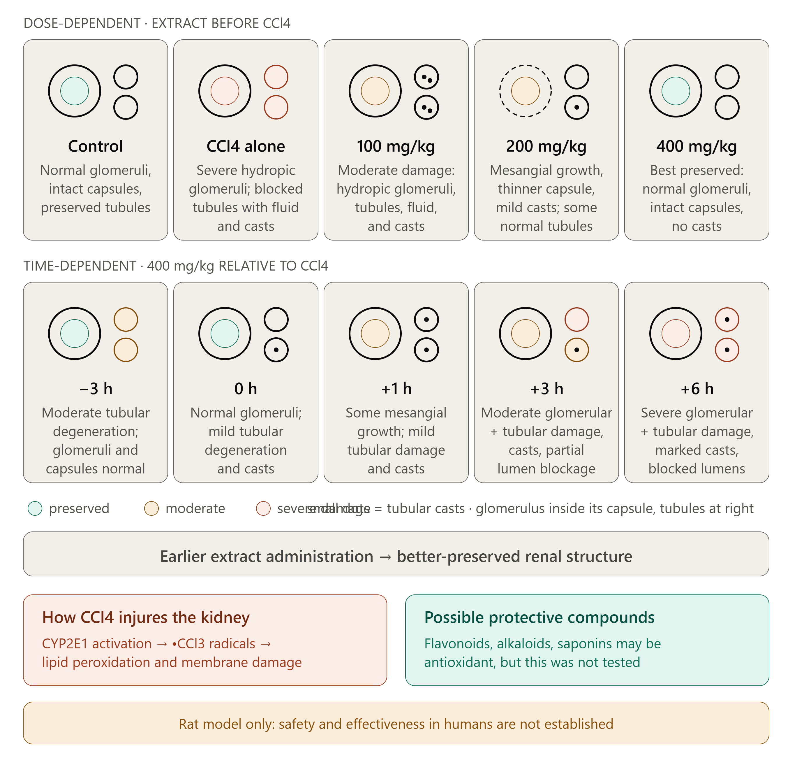
Animals receiving 100 mg/kg extract showed moderate histological alterations, including hydropic degeneration of the glomeruli and renal tubules. Tubular lumens also contained moderate amounts of fluid and casts.
At the 200 mg/kg dose, the histological examination revealed mesangial proliferation and thinning of Bowman's capsule, together with mild tubular cast deposition. However, areas of normal proximal and distal convoluted tubules remained visible.
The most favourable histological appearance was observed in animals pretreated with 400 mg/kg extract. Their kidney sections showed normal-looking glomeruli surrounded by intact Bowman's capsules, with no obvious tubular cast deposition.
These observations indicate that the highest tested dose was associated with the greatest degree of renal structural preservation.
3.3. Time-Dependent Effects of the Extract
The time-course experiment demonstrated that the timing of extract administration was another important factor in the observed protective response.
Histological examination revealed differences between animals receiving the 400 mg/kg dose at different intervals relative to CCl4 exposure.
Administration three hours before toxicant exposure was associated with moderate tubular degeneration, although the glomeruli and Bowman's capsules appeared normal.
When the extract was administered at zero hours, the kidney sections showed normal glomeruli and Bowman's capsules, with only mild tubular degeneration and tubular cast deposition.
Administration one hour after CCl4 exposure was associated with some degree of mesangial proliferation, mild hydropic tubular degeneration and mild tubular cast formation.
In contrast, administration three hours after toxicant exposure was associated with moderate glomerular degeneration, tubular degeneration, tubular cast formation and partial tubular lumen obliteration.
The most severe structural alterations were observed in the group receiving the extract six hours after CCl4 administration. These included severe glomerular degeneration, extensive hydropic tubular degeneration, marked tubular cast formation and tubular lumen obliteration.
Together, these observations suggest that the protective response was influenced by the interval between toxicant exposure and extract administration, with earlier administration associated with more favourable renal histological findings.
4. Histopathological Findings
The histological examination provided a visual assessment of the structural changes associated with CCl4-induced renal injury and the protective effects observed following treatment with the aqueous papaya seed extract.
The kidney sections from untreated control animals showed normal renal architecture, with clearly identifiable glomeruli, intact Bowman's capsules and preserved proximal and distal convoluted tubules.
Following CCl4 exposure, substantial structural damage became apparent. The glomeruli showed severe hydropic degeneration, while the proximal and distal tubular lumens were extensively obliterated and filled with fluid and casts.
In animals receiving the 100 mg/kg extract dose, moderate glomerular and tubular degeneration remained visible. The presence of fluid and casts within the tubular lumens indicated that renal tissue damage was still evident.
At 200 mg/kg, the kidney sections showed a more favourable appearance in certain areas, although mesangial proliferation, thinning of Bowman's capsule and mild tubular cast deposition were still observed.
The most preserved renal architecture was associated with the 400 mg/kg pretreatment group. The glomeruli and Bowman's capsules appeared normal, and no obvious tubular cast deposition was identified.
The time-dependent observations further demonstrated that delaying extract administration was associated with more pronounced structural alterations.
These findings complement the biochemical measurements and support the dose- and time-dependent nature of the nephroprotective response described in the study.
5. Discussion
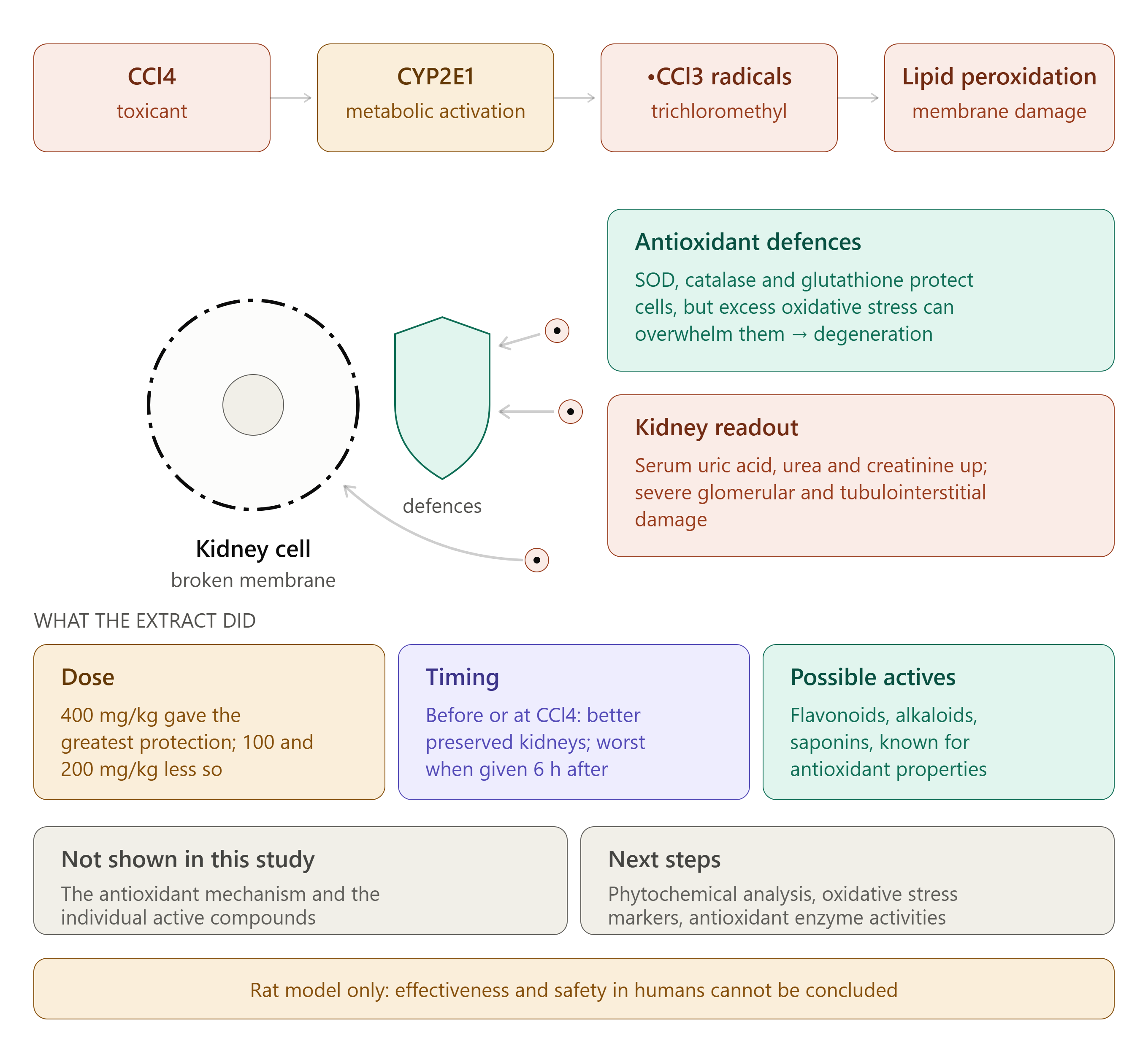
Carbon tetrachloride (CCl4) is widely used in experimental toxicology because of its ability to induce oxidative damage in biological tissues. Following metabolic activation by cytochrome P450 enzymes, particularly CYP2E1, it generates reactive trichloromethyl radicals (.CCl3), which promote lipid peroxidation and damage cellular membranes (Pohl et al., 1984; Fadhel and Amran, 2002; Basu, 2003).
Under normal conditions, antioxidant defence systems, including superoxide dismutase (SOD), catalase (CAT) and glutathione, help protect cells against reactive oxygen species. However, excessive oxidative stress can overwhelm these mechanisms, leading to cellular degeneration and tissue injury (Tom et al., 1984; Slater, 1984; Szymonik-Lesiuk et al., 2003).
In this study, CCl4 exposure caused significant increases in serum uric acid, urea and creatinine levels, indicating impaired renal function. These biochemical changes were accompanied by severe glomerular and tubulointerstitial damage, confirming the toxic effects of CCl4 on kidney tissues. Urea and creatinine are commonly used indicators for assessing renal function (Adelman et al., 1981).
5.1. Dose-Dependent Nephroprotective Effects
Pretreatment with aqueous Carica papaya seed extract reduced the biochemical alterations caused by CCl4. The protective response increased with the administered dose, with 400 mg/kg providing the greatest protection among the doses investigated.
Histological observations supported these findings. Moderate glomerular and tubular degeneration remained visible at 100 mg/kg, while the 200 mg/kg dose showed improved tissue appearance with some remaining alterations. The 400 mg/kg dose produced the most favourable results, with almost normal glomeruli and no obvious tubular cast deposition.
These findings suggest that the aqueous seed extract may help preserve renal structure and function under the experimental conditions of this study.
5.2. Influence of Treatment Timing
The timing of extract administration also influenced the observed protective effects. Animals receiving the 400 mg/kg dose before or at the time of CCl4 exposure generally showed better-preserved kidney structures than those treated several hours after exposure.
The most severe histological alterations were observed in animals receiving the extract six hours after toxicant administration. These findings suggest that earlier administration was associated with greater renal protection.
5.3. Possible Antioxidant Mechanisms
The protective activity of Carica papaya seed extract may be related to its phytochemical composition. Flavonoids, alkaloids and saponins are among the plant compounds that have attracted attention for their potential antioxidant and tissue-protective properties.
Previous studies have associated these compounds with antioxidant and protective activities (Miller and Rice-Evans, 1997; Jeong et al., 1996; Adeneye and Benebo, 2008). Their presence in the aqueous papaya seed extract (Adeneye and Olagunju, in press) suggests that they may contribute to the observed nephroprotective effects.
However, the present study did not directly investigate the antioxidant mechanisms or identify the individual compounds responsible for this activity. Further phytochemical and molecular investigations are therefore necessary.
5.4. Research Perspectives and Limitations
Although the results demonstrate promising protective effects in CCl4-treated rats, further studies are needed to identify the active phytochemical compounds and clarify their mechanisms of action. Additional investigations of oxidative stress markers and antioxidant enzyme activities could provide a better understanding of the biological effects.
Moreover, these findings are limited to an experimental animal model and cannot establish the effectiveness or safety of the extract in humans.
6. Conclusion
This study demonstrates the potential nephroprotective activity of aqueous Carica papaya seed extract against CCl4-induced renal injury in male Wistar rats.
The extract reduced biochemical indicators of renal damage and improved kidney tissue appearance, with the highest tested dose of 400 mg/kg showing the greatest protective effect. The timing of administration also influenced the observed response, with earlier treatment associated with better histological preservation.
These effects may be related to the antioxidant properties of phytochemical constituents such as flavonoids, alkaloids and saponins. Further research is required to identify the active compounds and establish their mechanisms of action, safety and potential applications.
7. References
The original bibliography should be retained without modification, including all author names, publication details, page numbers, DOIs and source links.
Original references: Abraham and Wilfred (1999); Adelman et al. (1981); Adeneye and Benebo (2008); Adeneye and Olagunju (in press); Basu (2003); Biod and Sirota (1948); Canadian Council of Animal Care (1984); Duke (1984); Fadhel and Amran (2002); Gill (1992); Jeong et al. (1996); Lu et al. (2002); Marsh et al. (1965); Miller and Rice-Evans (1997); Ogunyemi et al. (2008); Olagunju et al. (1995); Pohl et al. (1984); Slater (1984); Stal and Olson (2000); Szymonik-Lesiuk et al. (2003); Tom et al. (1984).